Data
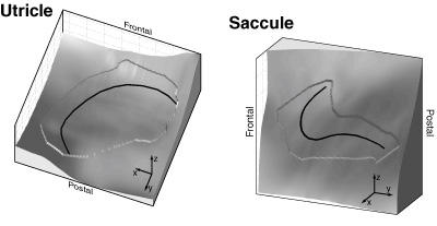
Utricle.mat (901 kB): "Utricle" is a structure, containing the {x / y / z}-data of the left utricle, in [mm]. The z-component of the utricle is eleveated by 0.05 mm with respect to the surrounding, and "indx" contains the locations of the edge. For technical reasons, the first and last column contains the slice number (or so ...). The coordinate system is described below.
Saccule.mat (811 kB): same as above, but with the structure "Saccule".
Methods
To determine hair cell polarization vectors across the epithelium,
we specified the curved surfaces of the utricular and saccular epithelia. They
were obtained from the measurements of Takagi et al. of human labyrinths (Takagi
et al., 1988). The original data set contained the geometrical locations,
organized in rows that corresponded to slices of the maculae. This data set
was pre-processed to remove artefacts of slicing. Single misplaced rows were
shifted or deleted to create smooth surfaces. Additional points in the periphery
of the measured data points were introduced which helped to smoothly extend
the macular surface beyond the original data set. The subsequent use of this
geometrical data required the representation of the surfaces on a regular grid.
A standard interpolation scheme was employed for this purpose. Remaining artefacts
of the slicing procedure were removed by smoothing the data set with a Savitzky-Golay
filter. Filter settings ensured that only artefacts were removed.
The geometrical data were represented with respect to a coordinate system that
allowed simple comparison with existing observations. We employed the same system
as used by Goldberg et al. (1976). In this coordinate
system, the x-axis points out the left ear, the y-axis out of the back of the
head and the z-axis out of its top. Since the original data set also included
the semi-circular canals, it was possible to use their orientation together
with the measured orientation of the semi-circular canals (Blanks
et al. 1975) to adjust the data set by a sequence of rotations.
Regions that represent measured data points are elevated. The insets illustrate
the orientation of the coordinate system.
Blanks RHI, Curthoys IS, Markham CH (1975) Planar relationships of the semicircular canals in man Acta Otolaryngol 80: 156-196
Fernandez C, Goldberg JM (1976a) Physiology of peripheral neurons innervating otolith organs of the squirrel monkey. I. Response to static tilts and to long-duration centrifugal force J Neurophysiol 39: 970-984
Takagi A, Sando I (1988) Computer-aided three-dimensional reconstruction and measurement of the vestibular end-organs Otolaryngol Head Neck Surg 98: 195-202